Sangolkar
Heart Care Center
Welcome to Sangolkar heart care center- a place for complete care for heart diseases


Clinic
Welcome to Sangolkar heart care center- a place for complete care for heart diseases. We are located near civil hospital, Sangli, Maharashtra. Dr. Ravindra Sangolkar is highly skilled, well trained and one of the best interventional cardiologist in Sangli.
Our aim is to provide quality services for all heart related ailments. Here we provide complete cardiac care with facilities like ECG, 2D Echocardiography, TMT, Angiography, Angioplasty, Pacemaker insertion, Advanced angioplasty with use of imaging (IVUS/OCT), Device closures for congenital heart defects (ASD/PDA), Balloon Mitral Valvotomy. We also provide services under various government schemes.

Dr. Ravindra Sangolkar
Dr Ravindra Ramhari Sangolkar completed his MBBS degree at Govt Medical
College, Miraj. He finished his MD Medicine at Govt Medical College, Latur. He then moved to Hyderabad for special training in Cardiology. He has worked as Consultant Cardiologist at Care hospitals, Hyderabad after finishing Cardiology training. During that time, he excelled in complex cardiac
interventions like Angioplasties, pacemaker insertion.
He has also published many papers in international journals and was also invited to give talk in international cardiology conference held at Bangkok, Thailand. He is currently working at Sangolkar Heart Care center, Sangli. He is also a panel cardiologist at various multispeciality hospitals like Synergy hospital, Bharti hospital and Kulloli hospital.
Youtube Channel – https://www.youtube.com/channel/UCHlpN5VDfDgEDlb_l4jGWGg


Specialities available at our clinic
2 D Echocardiography
हृदयप्रतिध्वनी आलेख चाचणी म्हणजेच हृदयाची अल्ट्रासोनोग्राफी ही तपासणी होय. यात हृदयाची रचना, कप्पे, झडपा, त्यांची कार्यक्षमता, त्यांचे आकारमान याबद्दल सविस्तर माहिती मिळते. त्यातच टू-डी (द्विमिती-two dimensional Echo) आणि कलर डॉप्लर (Colour Doppler) या उच्च दर्जाच्या चाचण्या उपलब्ध असून त्यांच्या मदतीने हृदयविकार ओळखणे हे सहजसाध्य झाले आहे आणि तंतोतंत रोगनिदान करून त्यावरील उपाययोजना कितपत परिणामकारक ठरले आहे याचे पण अवलोकन केले जाते.
या चाचणीत अल्ट्रासाऊंड ध्वनीलहरी या शरीरात सोडल्या जातात व त्या आतील अवयवापर्यंत धडकून, प्रवर्तित होऊन शरीराबाहेर, मायक्रोफोनसारख्या ‘ट्रान्सडय़ूसर’द्वारे पुन्हा शोषून संगणकाद्वारे त्याचे अवलोकन करून पडद्यावर चित्राच्या रूपात दाखवल्या जाते व त्याद्वारे हृदयाच्या विविध आजाराच्या कार्याची माहिती तज्ज्ञ डॉक्टरला मिळू शकते.
२-डी एको (2-D Echo- Two Dimentional Echo, द्विमिती इको) यामध्ये हृदयाचे आकुंचन प्रसरण पावणे, हृदयाची कार्यक्षमता, हृदयाच्या वेगवेगळय़ा झडपा आणि त्यांची उघडझाप या गोष्टींचे आकलन चलचित्राद्वारे तज्ज्ञांना करता येते.
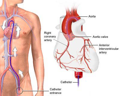
Angiography
अँजिओग्राफीमध्ये केवळ हृदयाच्या रक्तवाहिन्याची तपासणी केली जाते. अँजिओग्राफीत हृदयाच्या स्नायूला रक्तपुरवठा देणाऱ्या रक्तवाहिन्यांमध्ये (कोरोनरी आर्टरीज) गाठी निर्माण झाल्या आहेत का? ते पाहिले जाते. अँजिओग्राफीमुळे हृदयाच्या रक्तवाहिन्यांतील ब्लॉकेजची स्थिती कळण्यास मदत होते. ह्या तपासणीला coronary angiography किंवा cardiac angiogram ह्या नावानेही ओळखले जाते. आपले डॉक्टर एखाद्यास हार्ट अटॅक येण्याची शक्यता वाटत असल्यास ते त्याची अँजिओग्राफी करून पाहू शकतात. याशिवाय छातीत दुखणे, unstable angina, aortic stenosis, हार्ट फेल्युअर यामध्ये अँजिओग्राफी केली जाते.
View Speciality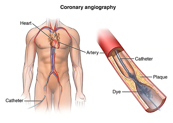
Angioplasty
कोरोनरी अँजिओग्राफी या तपासणीमध्ये जर कोरोनरी आर्टरीला अवरोध (Block) असल्याचे निदर्शनास आले. या अवरोधाची (Block) ट्रीटमेंट म्हणजे प्रत्येक वेळी बायपास सर्जरीच करावी लागते, असे नाही. गेल्या १५ वर्षांपासून कमी त्रासदायक, कमी विच्छेदन लागणारी औषधोपचार पद्धती (Treatment Modality) विकसित झाली असून तिचे नाव आहे. 'कोरोनरी अॅन्जिओप्लास्टी. यामध्ये कोरोनरी अँजिओग्राफी-प्रमाणेच मांडीच्या धमनीमधून गाइडिंग कॅथेटर (Guiding Catheter) नामक नळी कोरोनरी आर्टरीच्या मुखाजवळ ठेवण्यात येते. या Catheter मधून एक सूक्ष्म अशी वायर (Guiding Wire) पुढे सरकवतात. या वायरची हालचाल एक्स-रे स्क्रीनिंग मशीनमध्ये व्यवस्थित दिसते.आणि बाहेरून या वायरच्या सुरुवातीच्या भागाची हालचाल नियंत्रित करण्यात येते. अशाच नियंत्रित हालचाली करून हृदयरोगतज्ज्ञ ही वायर अवरोध असलेल्या आर्टरीमध्ये शेवटपर्यंत आत ढकलण्याचा प्रयत्न करतात जेणेकरून ही वायर अवरोधापलीकडे जाऊन स्थिरावेल. मग या वायरच्या साहाय्याने या वायरवरूनच डायलेटेशन कॅथेटरनामक एक छोटी नळी अवरोधापर्यंत (Block- पर्यंत) नेण्यात येते. या कॅथेटरच्या पुढील टोकास एक लांबूळका फुगा असतो (Baloon Dilation Catheter) एक्स-रे स्क्रीनिंगच्या साहाय्याने समोरच्या मॉनिटरवर बघून हा बलून अवरोध जिथे आहे, त्या योग्य ठिकाणी ठेवून त्याला फुगवण्यात येते. काही वेळानंतर मग फुग्यातील दाब कमी करण्यात येतो. असे दोन.. तीन वेळा करण्यात येते. अवरोध हा मुख्यत: चरबीने बनलेला असल्यामुळे या बलूनद्वारे दाबला जातो व आर्टरी पूर्ववत होऊ शकते. अवरोध हा मुख्यत: चरबीने बनलेला असल्यामुळे या बलूनद्वारे दाबला जातो व आर्टरी पूर्ववत होऊ शकते. मग वायर, बलून कॅथेटर आणि गाइडिंग कॅथेटर काढून घेण्यात येतात.
View Speciality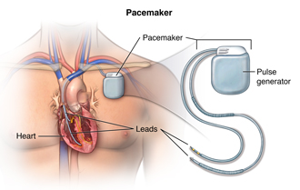
Pacemaker insertion
A pacemaker insertion is the implantation of a small electronic device that is usually placed in the chest (just below the collarbone) to help regulate slow electrical problems with the heart. A pacemaker may be recommended toensure that the heartbeat does not slow to a dangerously low rate.
View Speciality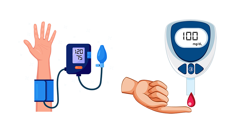
Blood pressure and Diabetes
management
High blood pressure can lead to many complications of diabetes, including diabetic eye disease and kidney disease, or make them worse. Most people with diabetes will eventually have high blood pressure, along with other heart and circulation problems.
View Speciality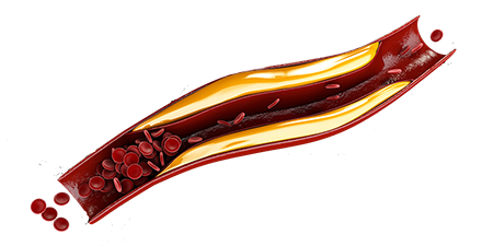
Cholesterol treatment
Cholesterol is a waxy substance found in your blood. Your body needs cholesterol to build healthy cells, but high levels of cholesterol can increase your risk of heart disease. With high cholesterol, you can develop fatty deposits in your blood vessels.
View Speciality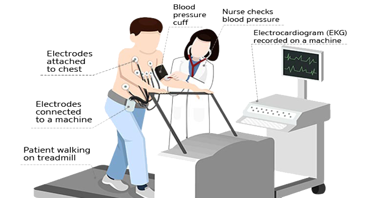
Treadmill test
ही साधी सरळ आणि सोपी तपासणी आहे. हृदयाला होणारा रक्तपुरवठा व्यवस्थित आहे की नाही, छातीचे दुखणे हे अन्जायना तर नाही ना, हे पाहण्यासाठी या चाचणीचा वापर केला जातो. या चाचणीत मनुष्य चालत असताना संगणकाद्वारे ईसीजी काढून रुग्णाचे रक्तदाब, हृदयाचे ठोके, त्याला होणारा त्रास, लागणारा दम किंवा इतर लक्षणे या चार गोष्टींची सांगड घालून हृदयविकाराचे निदान केले जाते.
View Speciality
Balloon mitral valvotomy
Balloon Mitral valvotomy successfully opens the narrowed valve
and improves the overall function of the heart. If balloon
valvotomy cannot be performed, surgical valve repair or
replacement may be options. Valve replacement (removing the
old valve and replacing it with a mechanical or biological valve) is
reserved for valves that are damaged beyond repair.
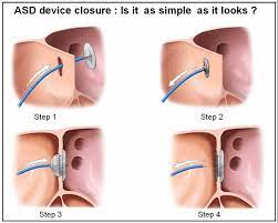
Device closure of congenitalbirth
defects (ASD/PDA)
The percutaneous closure of PFO and ASD is performed using a
special closure device. The device is folded or attached on to a
special catheter, similar to the catheter used during your catheterization. The catheter is inserted into a vein in the leg and
advanced into the heart and through the defect. The device is
slowly pushed out of the catheter allowing each side of the device
to open up and cover each side of the hole (like a sandwich),
closing the hole or defect. When the device is in proper position, it
is released from the special catheter. Over time, heart tissue grows over the implant, becoming part of the heart.

Thoughts from our best
patient’s experience
Seeking for verbal of our service quality? Find them here. Everything is transparent and straightforward for your sense of jusitifcation.
Best In Providing Treatment To The Required Diseases As Well As Understanding The Patients Mentality !!!!

Swapnil Khot
Patient
Hi all …..Initially I had lot of questions about my health symptoms. After meeting Dr Ravindra Sangolkar , I felt confident and motivated. Clinic and ambiance were clean and well maintained. Staff were kind and very polite and professional. And coming to billing, charges were very reasonable and pocket friendly.

Alisha Shaikh
Patient
With top notch experience and expertise , holistic approach Dr Sangolkars diagnosis and prognosis is precise. One of the best cardiologist and a great doctor.
Kiran Patil
Patient
Dr. Ravindra Sangolkar given the best heart treatment to me. Always available for consultation and suggestion. Proper advice and good helpline ECG. Thank you so much.
Yaseen Shaikh
Patient
Gallery
Youtube Channel – https://www.youtube.com/channel/UCHlpN5VDfDgEDlb_l4jGWGg